Protective Role of Lactobacillus acidophilus against vaginal infection with Trichomonas vaginalis
Abstract
Introduction: Vaginal colonization with lactobacilli species is characteristic of normal vaginal ecology. The absence of vaginal lactobacilli, particularly hydrogen peroxide producing isolates, has been associated with bacterial vaginosis and increased risk for sexually transmitted infection. In order to check the role of Lactobacillus acidophilus in vaginal infection when infected with Trichomonas vaginalis, in this study we mimic the vaginal condition in presence and absence of lactobacilli.
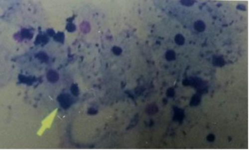
Methods: Forty Trichomonas vaginalis isolates from women referred to gynecology clinic were evaluated in this study. The parasites were isolated from vaginal discharge and urine in TYI-S-33media. The attachment of parasites, as pathogen, to vaginal epithelial cells was examined in healthy women in presence and absence of lactobacilli as well as their excretory-secretory products specially those of Lactobacillus acidophilus.
Results: Maximum number of vaginal epithelial cells infected by Trichomonas vaginalis was 37% (at 50 min from co-incubation) in experiments with parasites and vaginal epithelial cells (1st group) against 47% (at 40 min) in tube containing parasite along with Lactobacillus acidophilus and vaginal epithelial cells (2nd group). However, parasites in presence of vaginal epithelial cells and excretory-secretory product of Lactobacillus acidophilus (3rd group) showed the least number of infected cells 20% (at 40 min). Difference between 1st and 3rd group as well as 2nd and 3rd group of these experiments were significant (p<0.001). However, no significant difference were found between 1st and 2nd groups (p=0.28).
Conclusion: Modifiable biological and behavioral factors in vaginal ecology are associated to Lactobacillus colonization. This study suggests intervention strategies to improve vaginal health in women with risk of pathogensFull Text:
PDFReferences
Martin HL, Richardson BA, Nyange PM, Lavreys L, Hillier SL, Chohan B, et al. Vaginal lactobacilli, microbial flora, and risk of human immunodeficiency virus type 1 and sexually transmitted disease acquisition. The Journal of infectious diseases. 1999;180(6):1863-8.
Torok MR, Miller WC, Hobbs MM, Macdonald PD, Leone PA, Schwebke JR, et al. The association between Trichomonas vaginalis infection and level of vaginal lactobacilli, in nonpregnant women. The Journal of infectious diseases. 2007;196(7):1102-7.
Lepargneur JP, Rousseau V. [Protective role of the Doderlein flora]. Journal de gynecologie, obstetrique et biologie de la reproduction. 2002;31(5):485-94.
Valadkhan Z, Sharma S, Harjai K, Gupta I, Malla N. In vitro comparative kinetics of adhesive and haemolytic potential of T. vaginalis isolates from symptomatic and asymptomatic females. Indian journal of pathology & microbiology. 2003;46(4):693-9.
de Miguel N, Lustig G, Twu O, Chattopadhyay A, Wohlschlegel JA, Johnson PJ. Proteome analysis of the surface of Trichomonas vaginalis reveals novel proteins and strain-dependent differential expression. Molecular & cellular proteomics : MCP. 2010;9(7):1554-66.
Ryu JS, Min DY. Trichomonas vaginalis and trichomoniasis in the Republic of Korea. The Korean journal of parasitology. 2006;44(2):101-16.
Sobel JD. Vaginitis and vaginal flora: Controversies abound. Curr Opin Infect Dis. 1996;9(1):42-7.
Velraeds MM, van der Mei HC, Reid G, Busscher HJ. Inhibition of initial adhesion of uropathogenic Enterococcus faecalis by biosurfactants from Lactobacillus isolates. Applied and environmental microbiology. 1996;62(6):1958-63.
Valadkhani Z, Hassan N, Aghighi Z, Amirkhani A, Kazemi F, Esmaili I. The prevalence of Trichomoniasis in high risk behavior women attending the clinics of Tehran province penitentiaries. IJMS. 2010;35(3):190-4.
McLean NW, Rosenstein IJ. Characterisation and selection of a Lactobacillus species to re-colonise the vagina of women with recurrent bacterial vaginosis. J Med Microbiol. 2000;49(6):543-52.
Alderete JF, Demes P, Gombosova A, Valent M, Fabusova M, Janoska A, et al. Specific parasitism of purified vaginal epithelial cells by Trichomonas vaginalis. Infection and immunity. 1988;56(10):2558-62.
Wiesenfeld HC, Hillier SL, Krohn MA, Landers DV, Sweet RL. Bacterial vaginosis is a strong predictor of Neisseria gonorrhoeae and Chlamydia trachomatis infection. Clinical infectious diseases: an official publication of the Infectious Diseases Society of America. 2003;36(5):663-8.
Pi W, Ryu JS, Roh J. Lactobacillus acidophilus contributes to a healthy environment for vaginal epithelial cells. The Korean journal of parasitology. 2011;49(3):295-8.
A VS, E AK. Vaginal Protection by H2O2-Producing Lactobacilli. Jundishapur journal of microbiology. 2015;8(10):e22913.
Moreno-Brito V, Yanez-Gomez C, Meza-Cervantez P, Avila-Gonzalez L, Rodriguez MA, Ortega-Lopez J, et al. A Trichomonas vaginalis 120 kDa protein with identity to hydrogenosome pyruvate:ferredoxin oxidoreductase is a surface adhesin induced by iron. Cellular microbiology. 2005;7(2):245-58.
Kucknoor A, Mundodi V, Alderete JF. Trichomonas vaginalis adherence mediates differential gene expression in human vaginal epithelial cells. Cellular microbiology. 2005;7(6):887-97.
Martino JL, Vermund SH. Vaginal douching: evidence for risks or benefits to women's health. Epidemiologic reviews. 2002;24(2):109-24.
Newton ER, Piper JM, Shain RN, Perdue ST, Peairs W. Predictors of the vaginal microflora. American journal of obstetrics and gynecology. 2001;184(5):845-53; discussion 53-5.
Baeten JM, Hassan WM, Chohan V, Richardson BA, Mandaliya K, Ndinya-Achola JO, et al. Prospective study of correlates of vaginal Lactobacillus colonisation among high-risk HIV-1 seronegative women. Sex Transm Infect. 2009;85(5):348-53.
Juarez T, de L, de R, Nader-Macias ME. Estimation of vaginal probiotic lactobacilli growth parameters with the application of the Gompertz model. Canadian journal of microbiology. 2002;48(1):82-92.
Refbacks
- There are currently no refbacks.
