Morphological and crystallographic analysis of kidney stones in Eastern Morocco
Abstract
Abstract: Renal lithiasis is a common disease in the eastern region of Morocco, as it threatens the public health because of its repetitive nature as well as the complications it can cause in the kidney.
Objective: This study aims to investigate the morphological, molecular, and crystallographic characteristics of kidney stones affecting patients, specifically in the eastern region of Morocco.
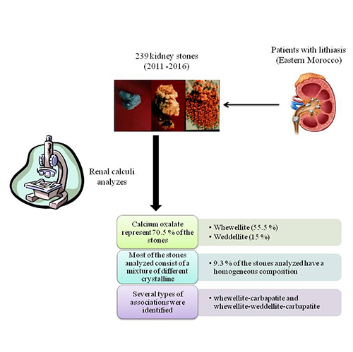
Methods: Morphological and constitutional analyses of 239 renal lithiases made it possible to identify the various crystalline forms present, to assess the structure of the stones and to determine the composition of their nucleation zone.
Results: It appears that calcium oxalate is the main component of the analyzed stones. It represents 70.5 % of the stones, which 55.5 % are majority whewellite and 15 % in weddellite. In comparison, calcium phosphates and magnesium (carbapatite and struvite) are the majority in only 8.9 % of cases, uric acid in 19.8 % of stones, and ammonium acid urate in 0.8 %. Calcium oxalate is predominant in the core of 52.5 % of the stones, carbapatite in 24.1 %, and uric acid in 20.3 % and struvite in 2.9 %. Most of the stones analyzed consist of a mixture of different crystalline constituents. Only 9.3 % of the stones analyzed have a homogeneous composition. Several types of associations were identified, the main ones being whewellite-carbapatite and whewellite-weddellite-carbapatite. Conclusion: The present study shows that calcium oxalate is the most common compound in the samples studied, followed by the uric acid compound.
Full Text:
PDFReferences
- T. Alelign, B. Petros, Kidney stone disease: an update on current concepts, Advances in urology, 2018, 1-12.
- M. López, B. Hoppe, History, epidemiology and regional diversities of urolithiasis, Pediatric Nephrology, 2010, 25, 49–59.
- A. Tiwari, V. Soni, V. Londhe, A. Bhandarkar, D. Bandawane, S. Nipate, An overview on potent indigenous herbs for urinary tract infirmity: Urolithiasis, Asian Journal of Pharmaceutical and Clinical Research, 2012, 5, 7–12.
- G. C. Curhan, Epidemiology of Stone Disease, Urol Clin North Am, 2007, 34, 287–293.
- A. Kamoun, M. Daudon, N. Kabaar, R. Dhaoui, S. Ben Ammar, Facteurs étiologiques de la lithiase urinaire de l’enfant en Tunisie, Progrès en urologie (Paris), 1995, 5, 942–945.
- D. Harrache, Z. Mesri, A. Addou, A. Semmoud, B. Lacour, Analyse des calculs urinaires de l’adulte dans l’Ouest Algérien par spectroscopie infrarouge à transformée de Fourier, L’Eurobiologiste (Paris), 1997, 31, 11–16.
- F. Meiouet, S. El Kabbaj, M. Daudon, Pediatric urolithiasis in Morocco: Composition of 432 urinary calculi analyzed by infrared spectroscopy, Progres en Urologie, 2019, 29, 173–182.
- M. Daudon, A. Valognes, C. Hennequin, P. Jungers, Importances de l’analyse morpho-constitutionnelle des calculs et des cristaux urinaires pour le diagnostic étiologique et le suivi thérapeutique de la maladie lithiasique, Spectra biologie, 1992, 5, 33–51.
- M. Daudon, R. J. Réveillaud, Typage morphologique des calculs oxalocalciques et données étiopathogéniques, Annales d’urologie, 1985, 19, 299–308.
- M. T. Keddis, A. D. Rule, Nephrolithiasis and loss of kidney function, Curr Opin Nephrol Hypertens, 2013, 22, 390–396.
- E. W. Vahlensieck, D. Bach, A. Hesse, A. Strenge, Epidemiology, pathogenesis and diagnosis of calcium oxalate urolithiasis, International urology and nephrology, 1982, 14, 333–347.
- M. Daudon, R. J. Réveillaud, L’analyse des calculs urinaires en routine importance pour le diagnostic étiologique de la lithiase, Fiches Pra Biol, 1985, 3, 43–49.
- M. Daudon, R. J. Réveillaud, Whewellite et weddellite: vers des éthiopathogénies différentes: intérêt dy typage morphologique des calculs, Néphrologie (Genève), 1984, 5, 195–201.
- M. Daudon, Plaidoyer pour une meilleure exploration des lithiases rénales, L’Eurobiologiste, 1997, 31, 17–35.
- P. Tosukhowong, C. Boonla, S. Ratchanon, M. Tanthanuch, K. Poonpirome, P. Supataravanich, T. Dissayabutra, K. Tungsanga, Crystalline composition and etiologic factors of kidney stone in Thailand: update 2007, Asian Biomedicine, 2007, 1, 87–95.
- M. L. Giannossi, G. Mongelli, F. Tateo, V. Summa, Mineralogical and morphological investigation of kidney stones of a Mediterranean region (Basilicata, Italy), Journal of X-Ray Science and Technology, 2012, 20, 175–186.
- H. Patel, Kidney Stones 2019: Epidemiology, Clinical Pathophysiology and Treatment, 2019.
https://hdl.handle.net/2152.5/6771.
- R. Z. Hossain, Y. Ogawa, S. Hokama, M. Morozumi, T. Hatano, Urolithiasis in Okinawa, Japan: A relatively high prevalence of uric acid stones, International Journal of Urology (2003), 2003, 10, 411–415.
- H. Bouzidi, D. de Brauwere, M. Daudon, Does urinary stone composition and morphology help for prediction of primary hyperparathyroidism? Nephrology Dialysis Transplantation, 2011, 26, 565–572.
- M. Daudon, Epidemiology of nephrolithiasis in France, Annales d’Urologie, 2005, 39, 209–231.
- P. Chatterjee, A. Chakraborty, A. K. Mukherjee, Phase composition and morphological characterization of human kidney stones using IR spectroscopy, scanning electron microscopy and X-ray Rietveld analysis, Spectrochimica Acta - Part A: Molecular and Biomolecular Spectroscopy, 2018, 200, 33–42.
- P. M. Patel, A. M. Kandabarow, A. Druck, S. Hart, R. H. Blackwell, A. Kadlec, A. Farooq, T. M. T. Turk, K. G. Baldea, Association of Impaired Renal Function With Changes in Urinary Mineral Excretion and Stone Composition, Urology, 2020. doi:10.1016/j.urology.2020.03.023.
- D. Harrache, Z. Mesri, A. Addou, A. Semmoud, B. Lacour, Analyse des calculs urinaires de l’adulte dans l’Ouest Algérien par spectroscopie infrarouge à transformée de Fourier, L’Eurobiologiste (Paris), 1997, 31, 11–16.
- R. C. Walton, J. P. Kavanagh, B. R. Heywood, P. N. Rao, Calcium oxalates grown in human urine under different batch conditions, Journal of crystal growth, 2005, 284, 517–529.
- N. K. Shapur, V. Uvarov, I. Popov, R. Katz, O. N. Gofrit, E. H. Landau, D. Pode, M. Duvdevani, Crystallite size - Is it a new predictor for renal stone burden?, Urology, 2012, 80, 980–985.
- A. Alaya, A. Nouri, M. Belgith, H. Saad, I. Hell, W. Hellara, R. Jouini, M. F. Najjar, Changes in kidney stones type according to sex and age in Tunisian patients, Actas Urológicas Españolas (English Edition), 2012, 36, 171–177.
- M. Daudon, O. Traxer, E. Lechevallier, C. Saussine, Epidemiology of urolithiasis, Progres en urologie: journal de l’Association francaise d’urologie et de la Societe francaise d’urologie, 2008, 18, 802-814.
- M. Daudon, Comment analyser un calcul et comment interpréter le résultat, L’Eurobiologiste (Paris), 1993, 27, 35–46.
- B. El Guerrouj, M. Bouhrim, Y. Bentata, M. Daudon, M. Melhaoui, L. Kharchoufa, N. Bencheikh, O. Bekkouch, Kidney Stone Disease (Urolithiasis): Epidemiological Study in the Eastern Region of Morocco, European Journal of Scientific Research, 2019, 155, 40–57.
- D. B. Leusmann, R. Blaschke, W. Schmandt, Results of 5035 stone analyses: a contribution to epidemiology of urinary stone disease, Scandinavian journal of urology and nephrology, 1990, 24, 205–210.
- T. Koide, T. Oka, M. Takaha, T. Sonoda, Urinary tract stone disease in modern Japan, European urology, 1986, 12, 403–407.
DOI: http://dx.doi.org/10.13171/mjc10502005261409hi
Refbacks
- There are currently no refbacks.
Copyright (c) 2020 Mediterranean Journal of Chemistry